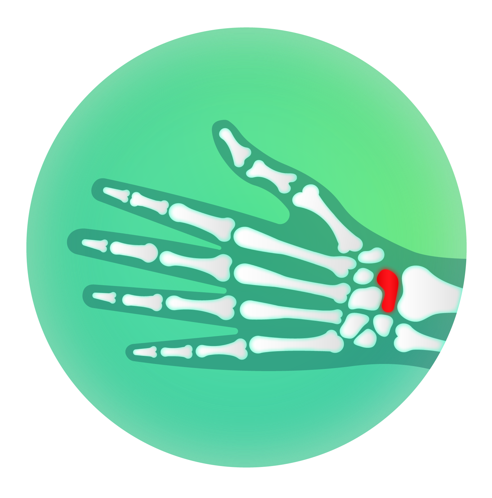
ScaphX
AI detection of scaphoid fractures in X-rays.
Modality:
X-ray
Pathology:
Occult scaphoid fracture
Status:
Current
MRI imaging is superior in identification of occult carpal fracture, but is not always accessible. Imaging from X-rays can give suboptimal views, and the presentation of arthritis can make small fractures difficult to see. An AI tool to aid clinical diagnosis of occult carpal fractures using x-rays would increase diagnostic sensitivity in areas and situations where MRI is not available.
A computer aided diagnosis tool which would automatically run when either a scaphoid fracture is suspected or if a patient is referred for a hand/wrist x-ray from A&E would increase sensitivity and confidence of diagnosis. Carpal fractures can be difficult to identify and patients with high clinical suspicion are put in a splint and referred to the fracture clinic even if a fracture isn’t seen on the x-ray by the clinician. Subtle lucency of an un-displaced fracture and the significance of a small bone fragment is currently easily missed. A successful tool would therefore increase diagnostic confidence and accuracy and reduce repeated x-rays and needless fracture clinic referrals
Clinical lead: Davina Mak
Patient pathway
A&E attendance with a suspected scaphoid fracture or hand/wrist injury.
Training data
Training data for ScaphX consists of 3 separate datasets - a set of x-rays of the wrist used to train the scaphoid classifier which locates the scaphoid bone in the x-ray; of approximately 2000 Xrays which had follow up MRIs within two weeks of the injury used to reach definitive diagnosis; and of approximately 8000 Xrays which do not have a corresponding MRI.
Testing data
The testing data for ScaphX consists of approximately 1000 Xrays which are not a part of the training dataset and are held separately.
Risks
The biggest risks is a negative disruption to the clinical pathway if there are too many false positive results which lead to needless treatment.
Goals
The reduction of need for further imaging where fractures can be diagnosed from Xrays alone, improving the patient experience and reducing waste.
Success criteria
A reduction in negative wrist MRIs.
Alternatives
Commercial applications were considered to solve this clinical problem but were found to be optimised for negatives while the clinical pathway at GSTT can only benefit from an AI tool which is optimised for positive scans.
Additional resources
TOHETI trial results
A critical review of AI use in scaphoid fracture detection
