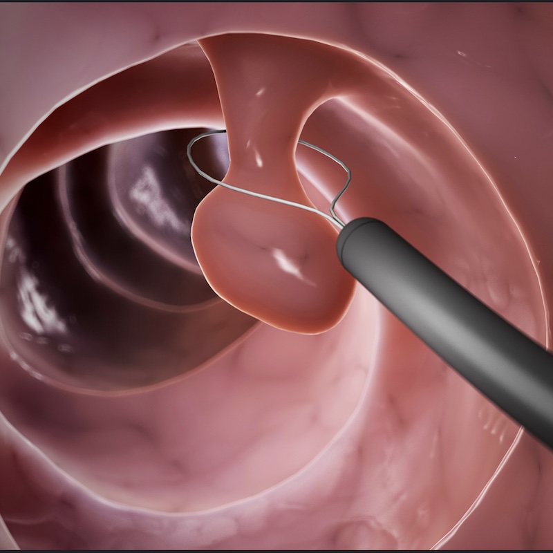
Detection of Rectal Cancer
AI detection of rectal cancers/polyps in MRI.
Modality:
MRI
Pathology:
Rectal cancer/polyps
Status:
Concluded

CSC Lead: Laurence
This project has been withdrawn. This decision was made due to lack of clinical time and complexity of 3D data. It may be revisited in the future.
MRI pelvis studies are reported by a variety of subspecialty radiologists leading to missed pathology in the rectum outside of the reporter’s immediate comfort zone. The consequence of a missed polyp is the development of later stage cancerous lesions which are more symptomatic and potentially incurable or require extensive surgery/treatment as opposed to minimally invasive endoluminal procedures. An artificially intelligent algorithm has the potential to reduce these errors by highlighting areas of concern particularly within the rectum which can be a challenging area to assess.
Clinical lead: Tina Mistry
Rationale
Development of an AI tool to identify areas of concern that may indicate early development of cancer and polyps in rectal MRI images
Patient pathway
Once a lesion is identified whether these abnormalities are cancerous or polyps the ultimate treatment for this is removal which is curable if performed early.
Training data
Large MRI pelvis dataset within the trust with the T2 weighted axial and diffusion performed in most cases. This will give a high yield of normal. Rectal cancer datasets also collected via a recent audit.
Risks
False positives will either result in early interval repeat imaging or proctoscopy performed by surgeons in clinic/endoscopy. False negatives may result in cancers being detected at a later stage.
Goals
An application that is able to identify areas of concern for further analysis by radiologists
Success criteria
Ability to identify lesions incidentally/early leading to improved outcomes for patients, early detection leads to stopping or reducing the development of malignancy which can be fatal when diagnosed at a late stage.
Alternatives
Currently no commercial products identified
References
Reducing the model variance of a rectal cancer segmentation network.