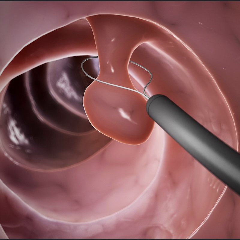
Early detection of rectal cancer
An application that identifies areas of concern on MRI images for the detection of rectal cancers/polyps
Modality:
MRI
Pathology:
Rectal cancer/polyps
Status:
Concluded

CSC Lead: Laurence
MRI pelvis studies are reported by a variety of subspecialty radiologists leading to missed pathology in the rectum outside of the reporter’s immediate comfort zone. The consequence of a missed polyp is the development of later stage cancerous lesions which are more symptomatic and potentially incurable or require extensive surgery/treatment as oppose to minimally invasive endoluminal procedures. An artificial intelligent algorithm has the potential to reduce these errors by highlighting areas of concern particularly within the rectum which can be a challenging area to assess.
Clinical lead: Tina Mistry
Project Plan
1. Meeting of all persons involved to determine AI specifications
**Setting technical and system requirements for AI model**
1. Agree on most useful metrics for success – e.g. percentage reduction in missed polyps. This will require designing a fixed methodology for measuring currently missed polyps which can be performed before and after model deployment.
2. Agree on minimum valuable requirements for success e.g. 25% reduction in missed polyps. We are aiming for a 10% reduction in missed polyps using the methodology above.
3. Agree on size of POC and production datasets and who is responsible for compiling them. We will use existing database of 5 year audit to generate proof-of-concept dataset
4. Agree on how model will be deployed. The application will push information back to radiologist workstation to indicate suspect dicom series as well as slice number and region
Development phase (proof of concept (POC)
- Dataset curation (retrospective)
- Model training
- Produce effective POC model
- Model testing
- Test model against agreed criteria for success (where applicable)
- Agreement to continue to production phase OR iterative/improve model OR discontinue
Development phase (production)
- Dataset curation (retrospective). Compile larger production dataset with all appropriate data properties (e.g. ethnicity, weight etc)
- Model training. Produce effective deployment model
- Model testing
- Test model against agreed criteria for success (where applicable).
- Clinical approval for model to be deployed
Deployment phase
- Deployment to test environment (if applicable)
- Deployment to hospital systems including training for users and continuous monitoring of model performance
References
Lee, Joohyung, et al. “Reducing the model variance of a rectal cancer segmentation network.” IEEE Access 7 (2019): 182725-182733. online