Lung Cancer Radiomics AI
AI to predict outcome in lung cancer.
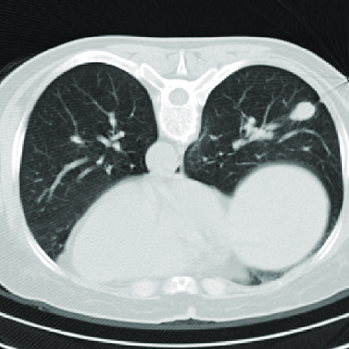
Modality:
Thorax CT (contrast enhanced)
Pathology:
Lung cancer with nodal involvement
Status:
Concluded
CSC Lead: Anil
This project has been withdrawn due to limited clinical capacity. This project may be revisited at a later date but it is not currently being actively worke on.
The survival rates from non-small-cell lung cancer is associated with TNM grading system (tumour, nodal involvement and metastatic extent), and also protein expression, however the only way to evaluate both is using histology results from an invasive biopsy investigation. Biopsies are also limited in that they may not fully characterise the tumour and spread of disease. The aim of this project is to use AI to extract RFs from the lymph nodes and lung cancer and correlate them with histology, to assess the RF ability to predict histology and patient outcomes. This will result in a non-invasive tool to build multivariate predictive models and stratify patient treatments.
Clinical lead(s): TBC
Rationale
Radiomic Features (RF) that are extracted from contrast enhanced CT of lung cancer and lymph nodes can be correlated with histology and survival outcomes to provide a non-invasive predictive tool to develop a multi-parametric predictive model to assist the decisions in patient treatment.
Patient pathway
Patients receive a contrast enhanced CT then a biopsy for diagnosis and staging of lung cancer, the results of which determine the treatment path.
Training data
Pre-operative Contrast enhanced Thorax CTs (2014-2018), collected into a database (n = 500 ~ 600). Option of including recent CTs (2018 – present) with limited data on survival after surgery.
Risks
Poor RF will develop a model with little predictive value. Mismanagement of patient treatment. Poor research outcomes.
Goals
A tool that extracts radiomic features with a high predictive power in predicting histology and patient outcomes.
Success criteria
Well-segmented primary lung lesions and intrathoracic lymph nodes. This will require clinicians’ supervision and validation against manual segmentation. Also, based on this, subsequent important criteria for AI success within this project will be the ability of RF automatically extracted from images to show correlation with histological parameters and outcome data.
Alternatives
Currently no commercial products identified.
References
Wang et al 2020
Ninatti et al 2020
Bashir et al 2019
Xu et al 2019
Lambin et al 2012
Wilson et al 2017
Xia et al 2019
Weikert et al 2019
Tsitsias et al 2020