Smart brain MRI protocols
Evaluation of brain MRI protocoling AI.
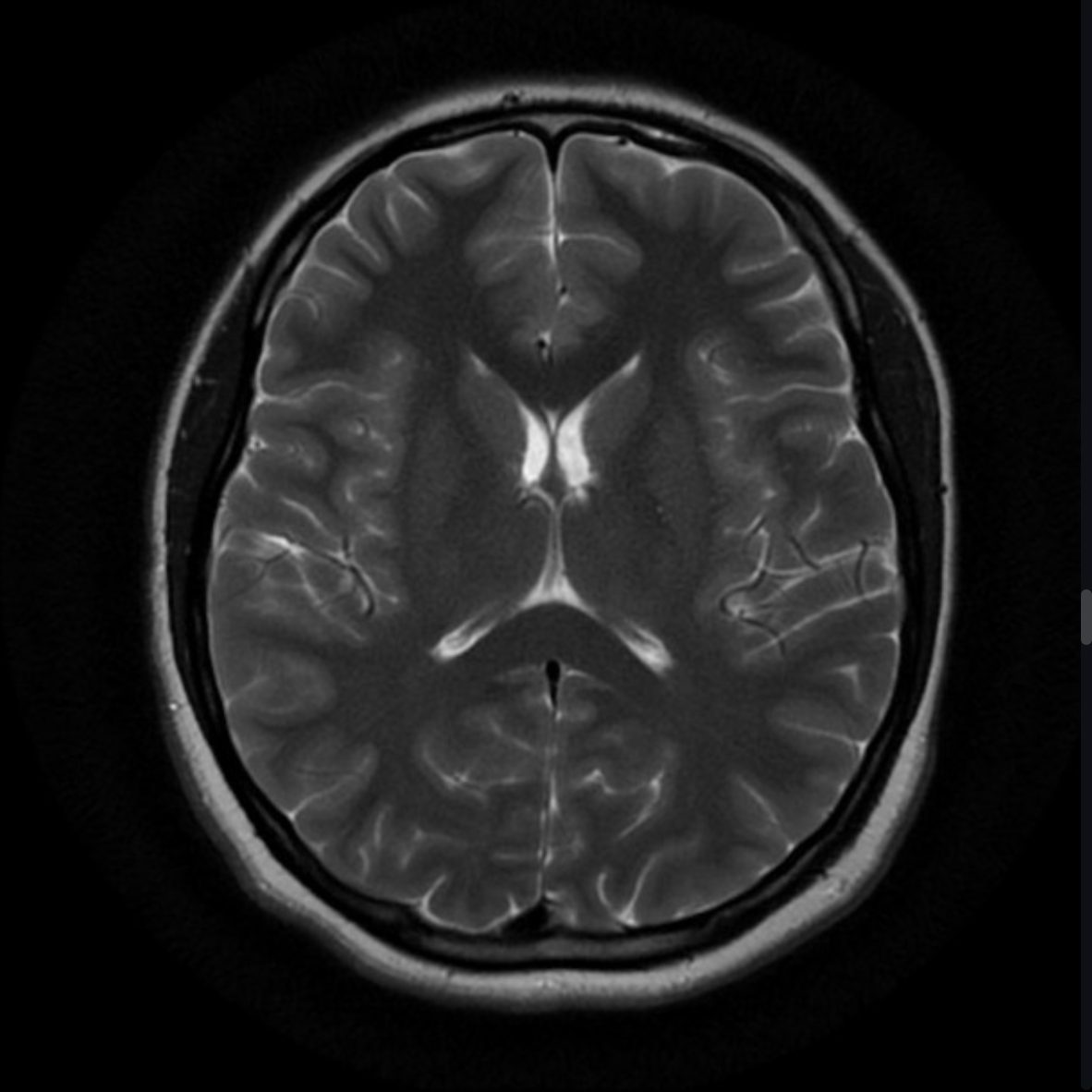
Modality:
MRI
Pathology:
Brain abnormalities
Status:
Concluded
CSC Lead: Laurence
This project has been completed. No further actions are planned on this topic.
A consequence of the ever-increasing demand for diagnostic MR imaging is additional workload and stress placed on MRI departments. MRI departments need to operate as efficiently as possible. A smart protocoling application for brain MRI data has the potential to improve the efficiency of the clinical MRI service and improve patient prioritisation.
Clinical leads: Asif Mazumder
Rationale
This application may also reduce scan time, re-examination rates and non-acute admission to ward environments.
Patient pathway
Patients who present to their GP with diffuse neurological symptoms (headaches, dizziness) are referred to GSTT for brain MRI. In current clinical practice, a radiologist will review these images and if an infarct is spotted, the radiologist must recall the patient for additional MRI scans. Instead, with a smart protocol in place, the infarct can be flagged in real-time for the attention of the radiologist and additional follow-up scans can be completed in the same appointment slot.
Testing data
Approximately a total of 5000 MRIs were used in this retrospective study, accounting for a range of possible neurological findings.
Risks
False negatives present the largest risks, potentially leading to missed diagnosis.
Goals
Reduction in dedicated image capture of healthy patients, leading to a reduction in reporting lists.
Success criteria
High sensitivity demonstrated to all pathologies. Rate of false negatives must be fewer than 2%.