3D Fetal Brain MRI MoCo
SaMD for brain reconstruction of fetal MRIs.
Modality:
MRI
Pathology:
Congenital birth defects
Status:
Concluded
CSC Lead: Tom
This project has been withdrawn due to issues with local deployment of the software.
Rationale
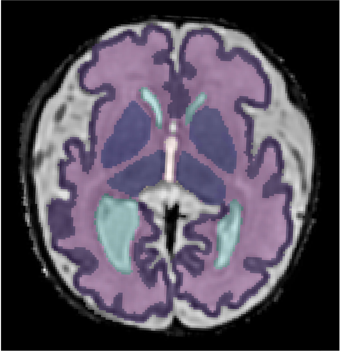
Fetal MRI is predominantly performed using 2D MRI, but this approach is susceptible to motion artefacts when the baby moves. Motion-corrected 3D MRI produces higher quality images, with the potential to improve diagnosis of abnormalities.
Academic Partners
Mary Rutherford, Joseph Hajnal, Maria Deprez, Alena Uus @ Perinatal Imaging & Health, King’s College London
Project Overview
Fetal MRI is an important adjunct to ultrasound imaging as it can provide additional confidence and insights when making a prenatal diagnosis. However, fetal motion during MRI scans can cause images to be blurry and not of sufficient diagnostic quality. In some cases, fetal motion can be so severe that the radiographer has to reacquire multiple scans until quality images are obtained, which can be very inefficient.
For more than 15 years, researchers in the department of Perinatal Imaging & Health at King’s College London have been developing advanced motion-correction MRI techniques. These techniques can provide artefact-free 3D MRI reconstructions of the fetal brain, heart and body.
In this project, CSC is working closely with the team at KCL to make these advanced 3D MRI techniques easier and more efficient for clinicians to use, and to make it easier for other hospitals to adopt these tools in their clinical pathways. For example, one of the first apps available on AIDE will be for motion-corrected 3D fetal brain MRI.
3D fetal brain MRI is performed by reconstruction of single-slice 2D MRI images into 3D volumes using slice-to-volume registration (SVR – www.svrtk.github.io) methods. Now, with the advent of machine learning technologies, it is possible for hospitals to perform 3D fetal brain MRI in a fully automated manner.